Histopathology

21 Gestational Weeks Inferior Hippocampal Formation colabeled with Calbindin, PCP4 and Calretinin
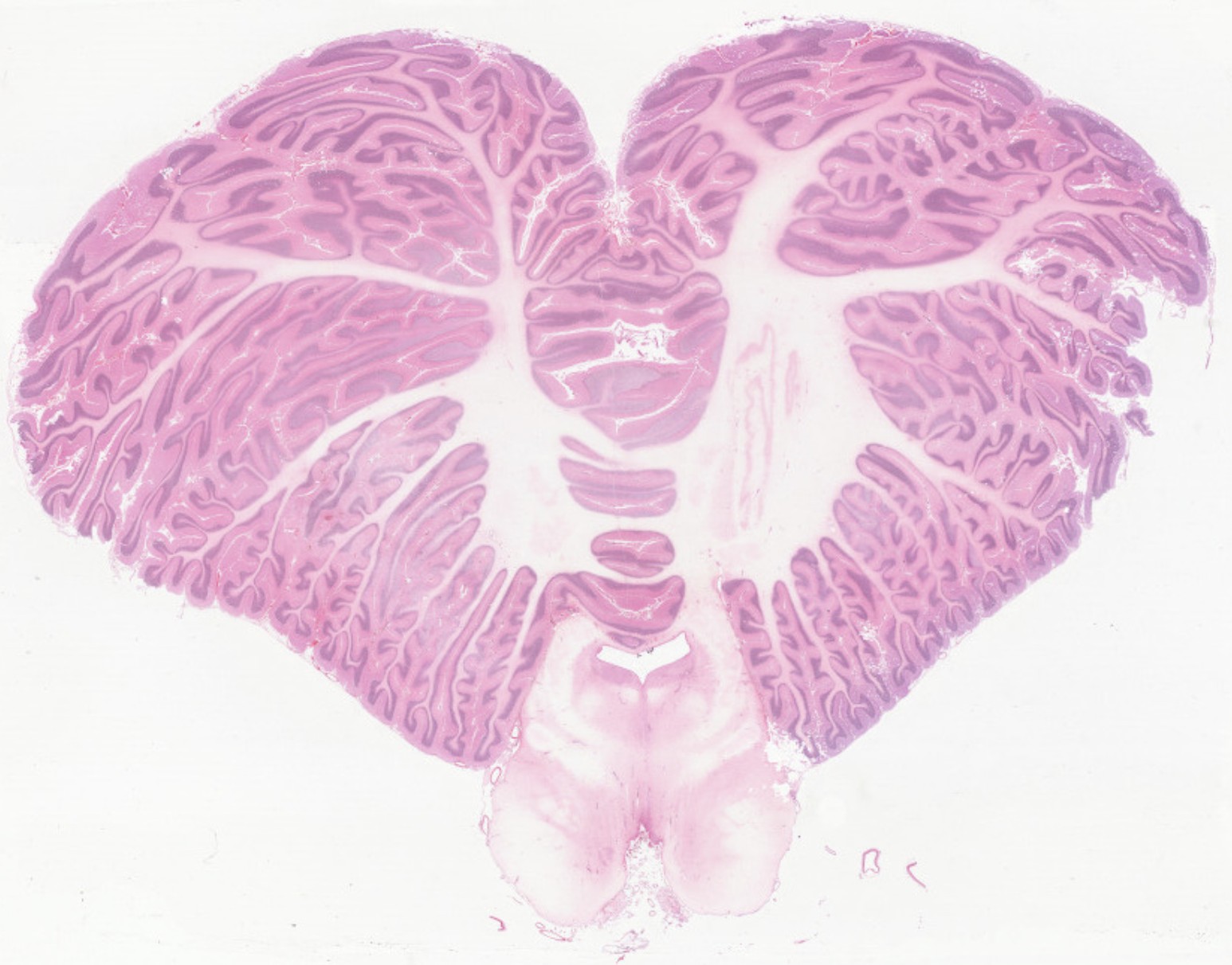
Adult Human Brain Cerebellum H&E stain
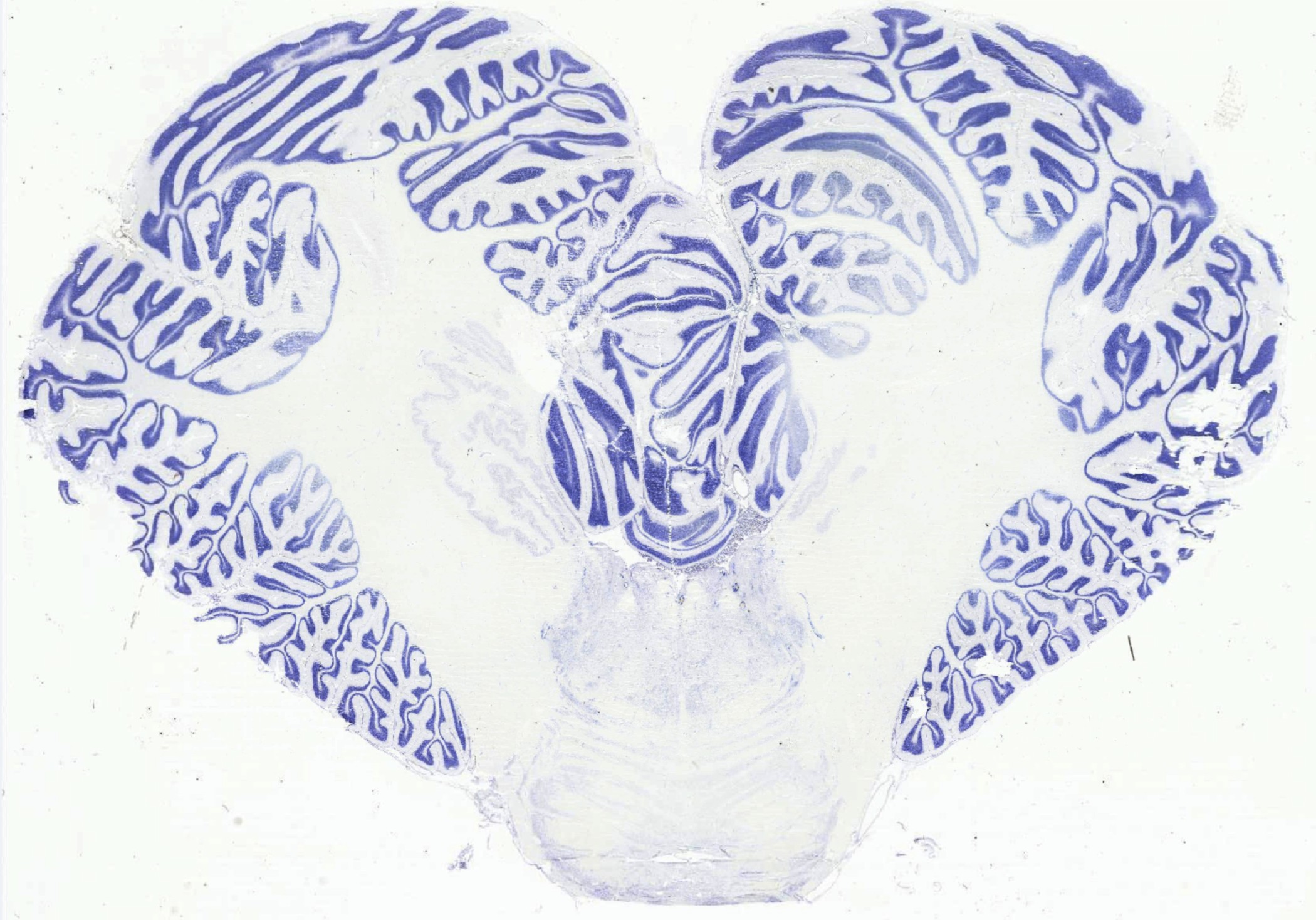
Adult Human Brain Cerebellum Nissil stain
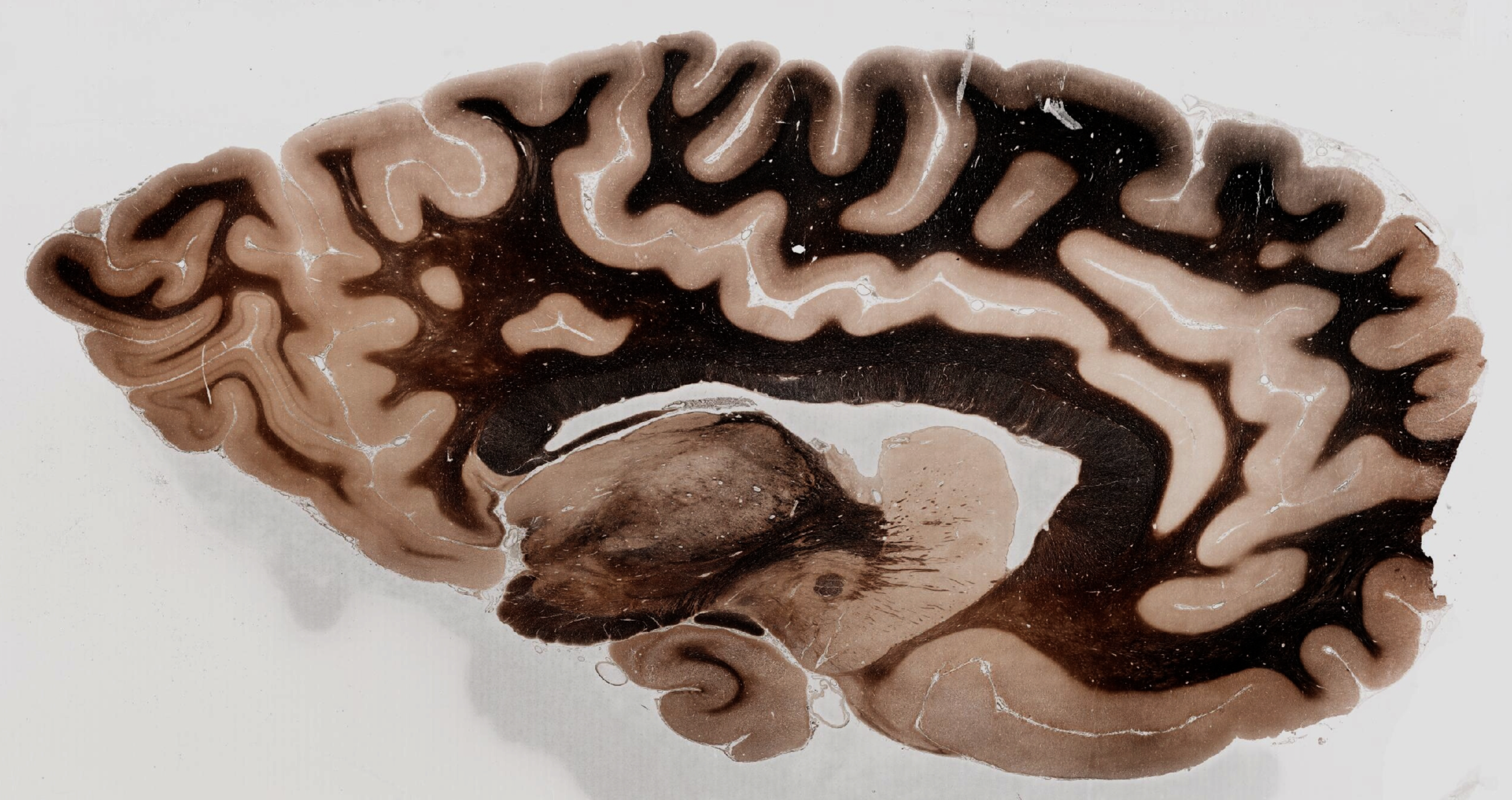
Adult Human Brain Myelin Stain
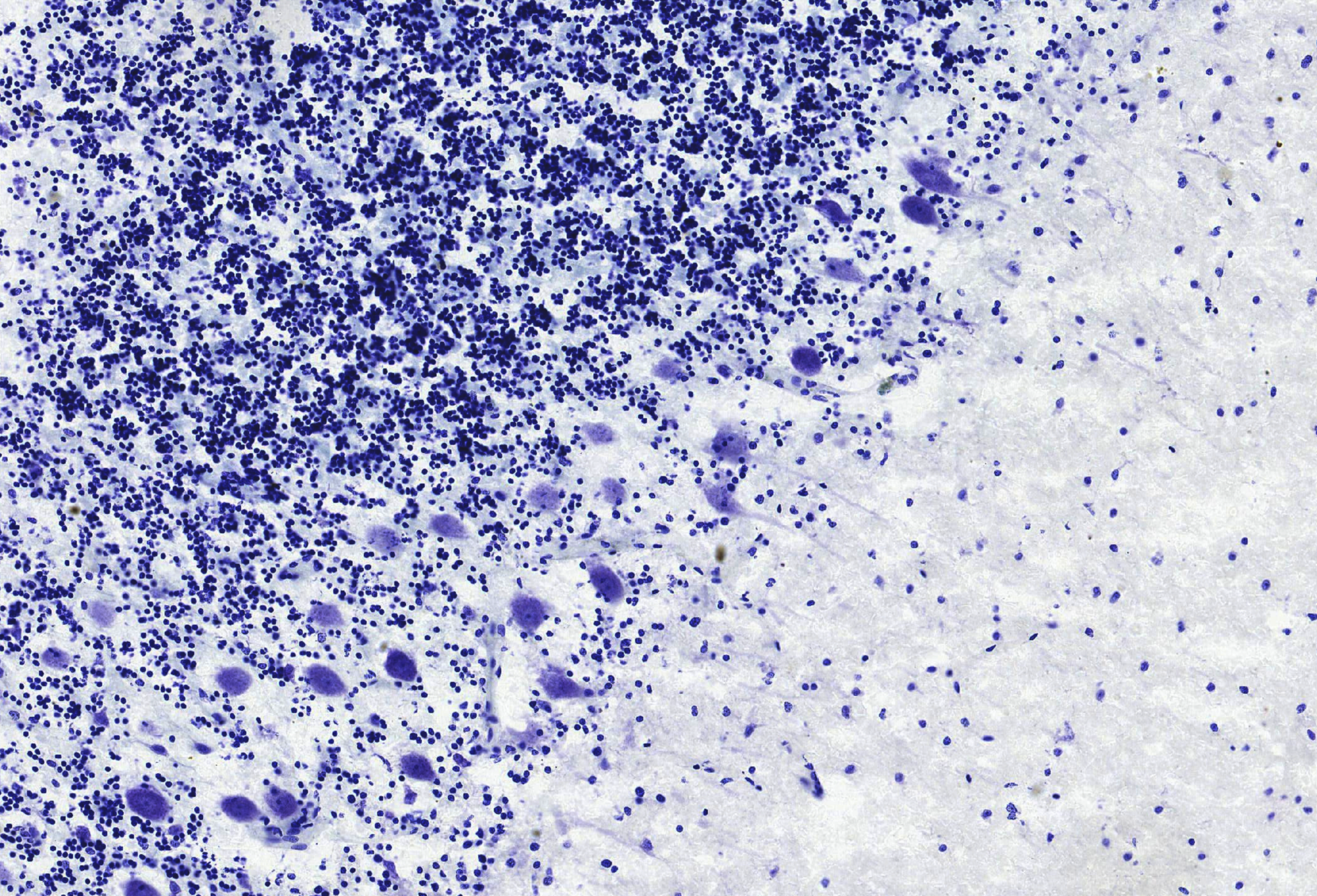
Adult Human Brain- Cerebellar Purkinje Cells

Adult Human Brainstem Immunostained for Neurofilament H
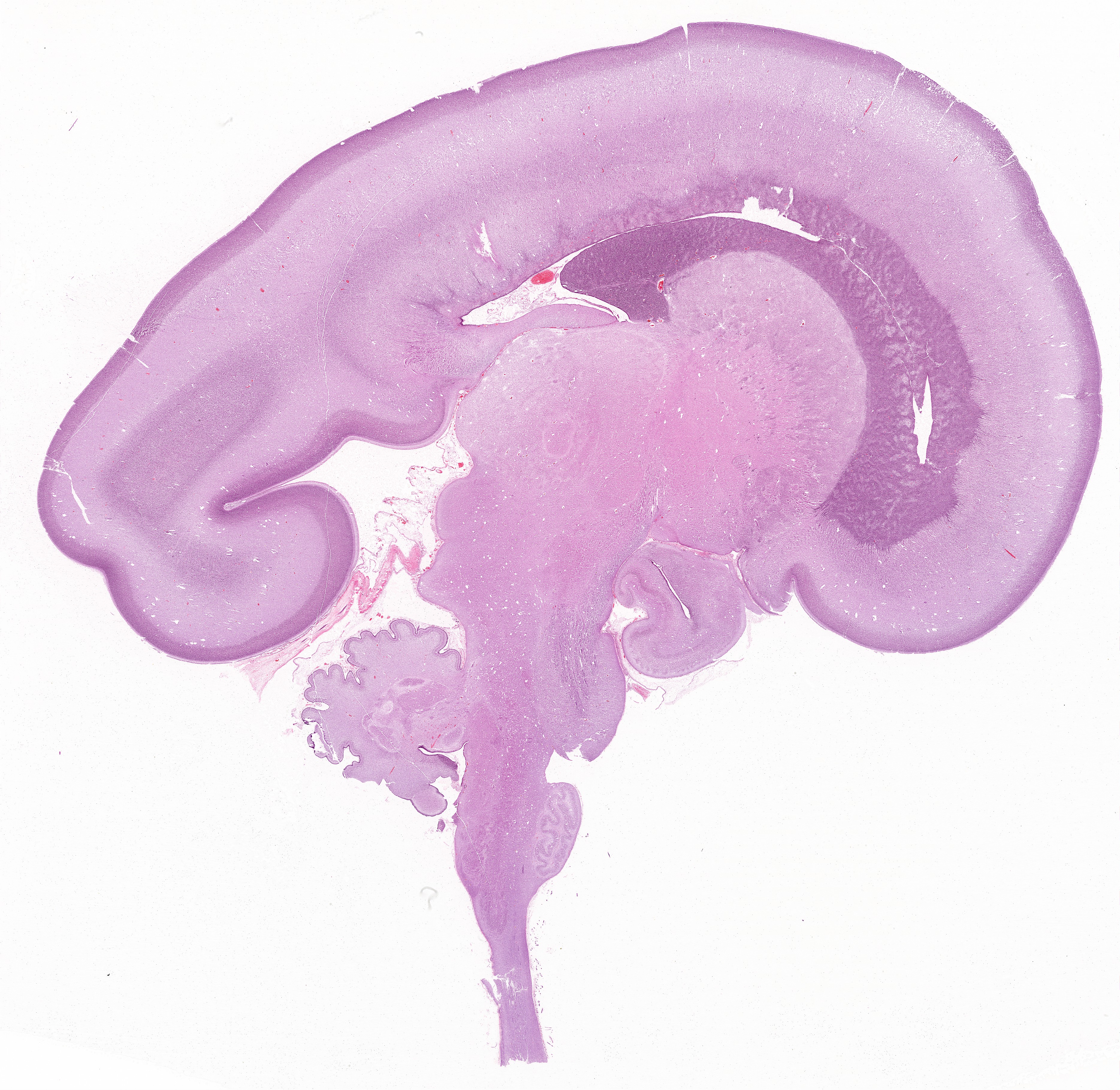
Fetal Brain H&E stained

Fetal Brain Hippocampal Formation Nissil Stain
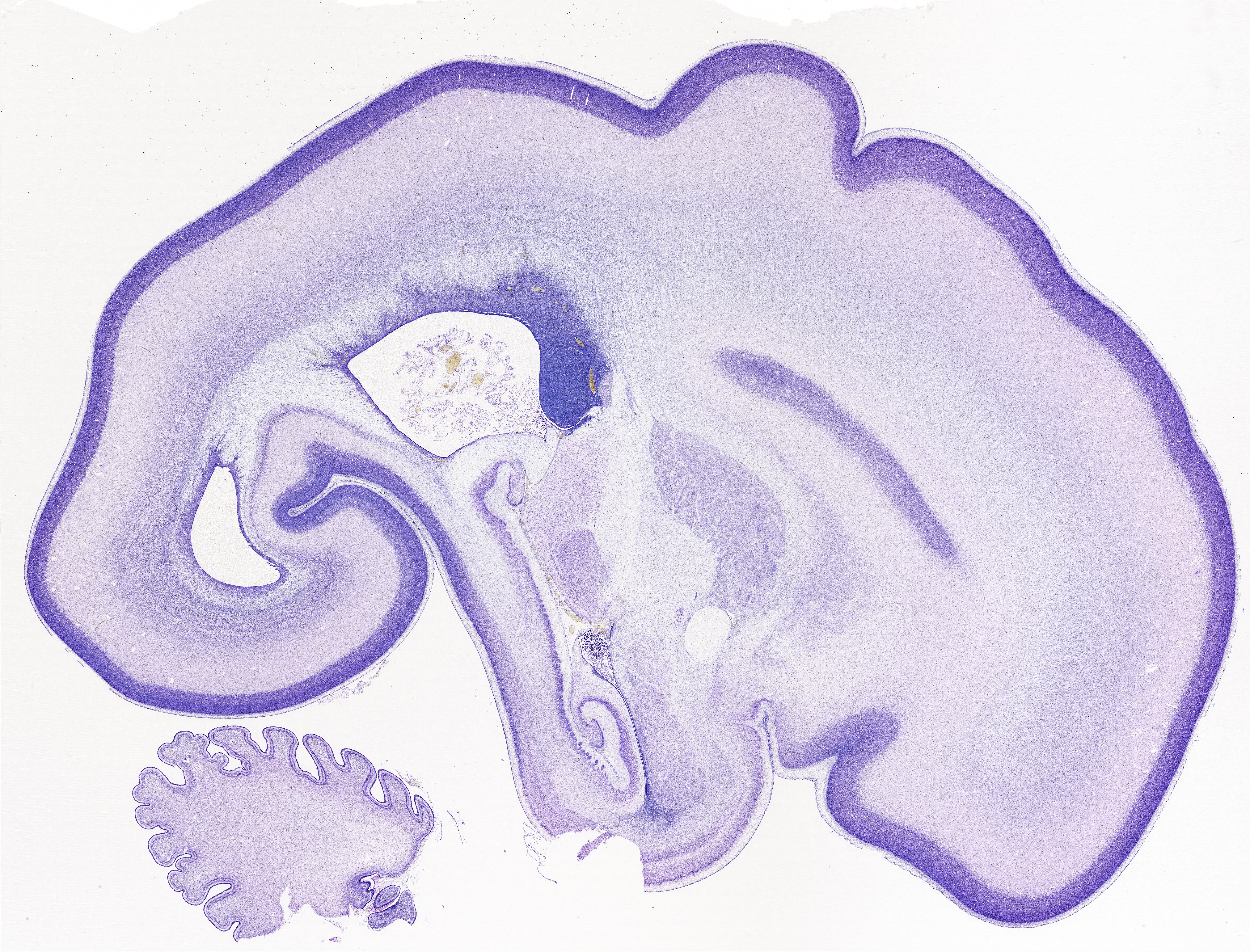
Fetal Brain Nissil Stain
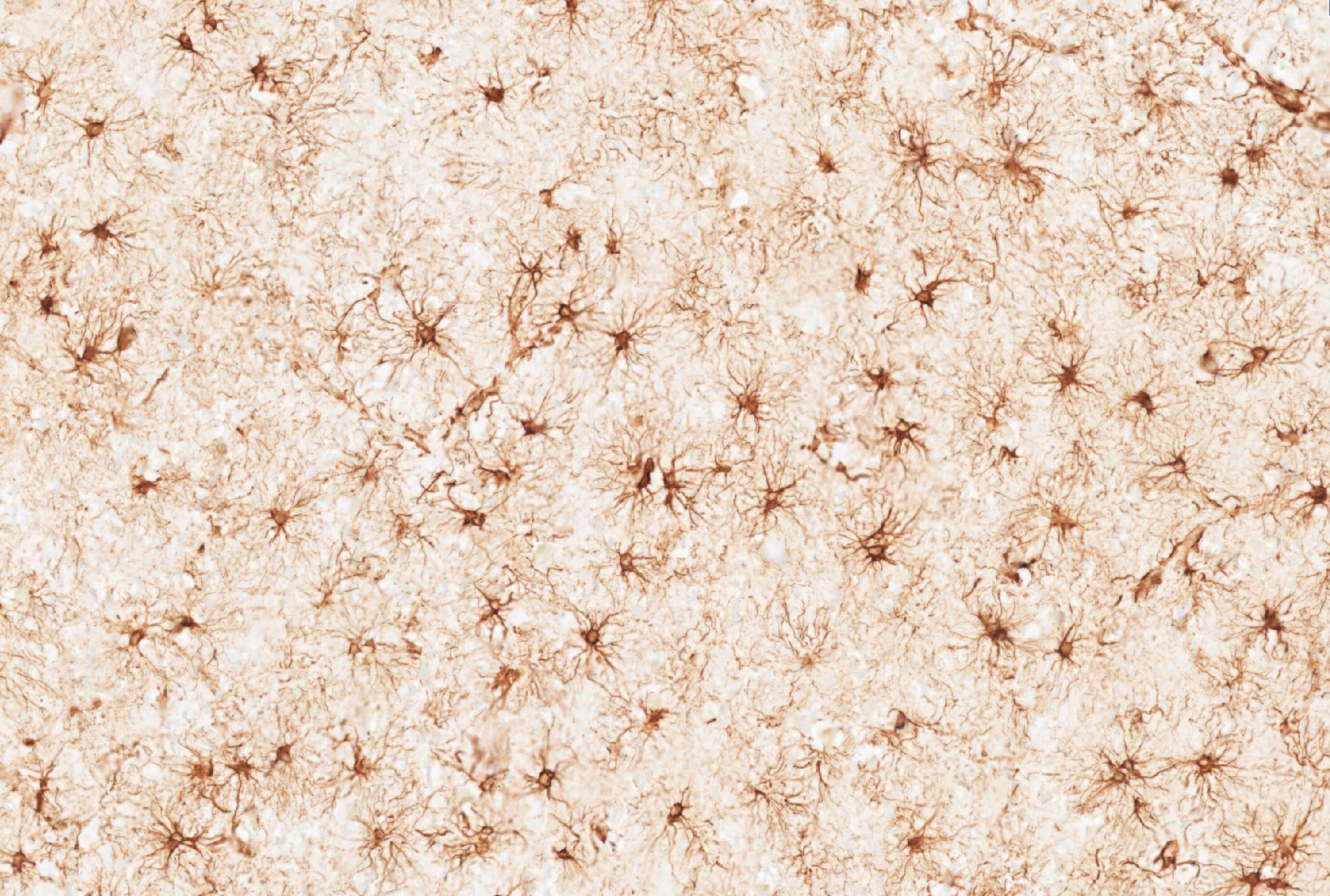
Reactive astrocytes in Ischemic stroke - GFAP IHC
Computational
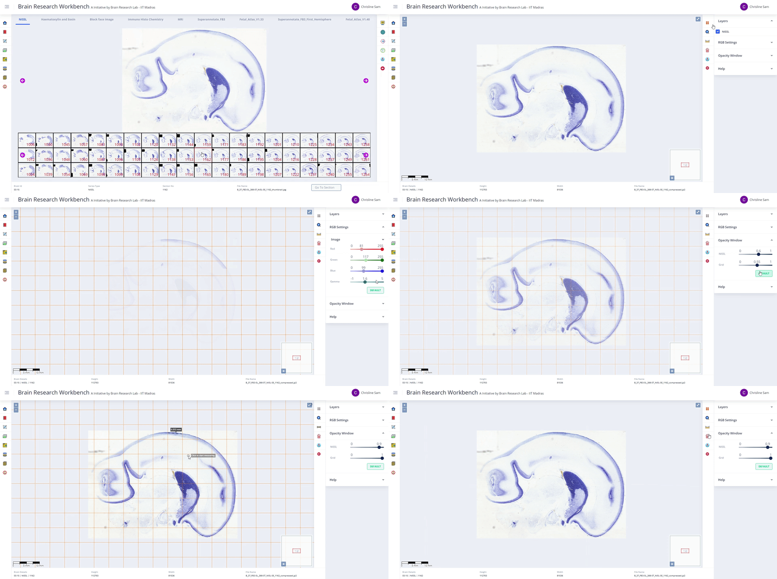
Zero Footprint Viewer
This tool enables viewing of microscopically scanned brain images at ultra-high resolution without loss of quality. It supports visualisation of 20 µm brain sections scanned at resolutions as fine as 0.5 µm.
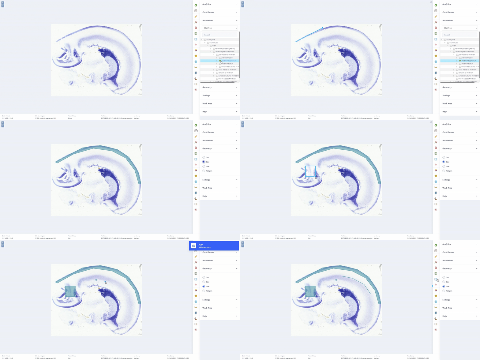
Atlas Creator
This tool supports geometry-based annotation of regions on high-resolution microscopic brain images. It allows creation, saving, display, and sharing of multiple region-specific annotations across users.
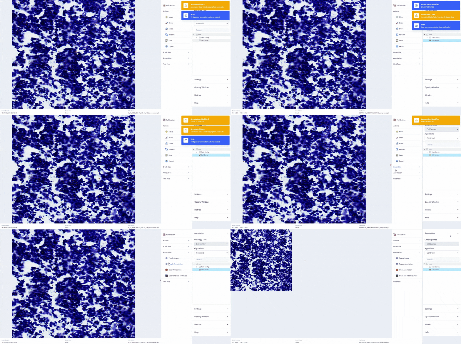
Cell Annotation Tool
This tool enables pixel-level manual annotation of cells on brain images. It supports machine-learning–based cell detection through high-quality training annotations.
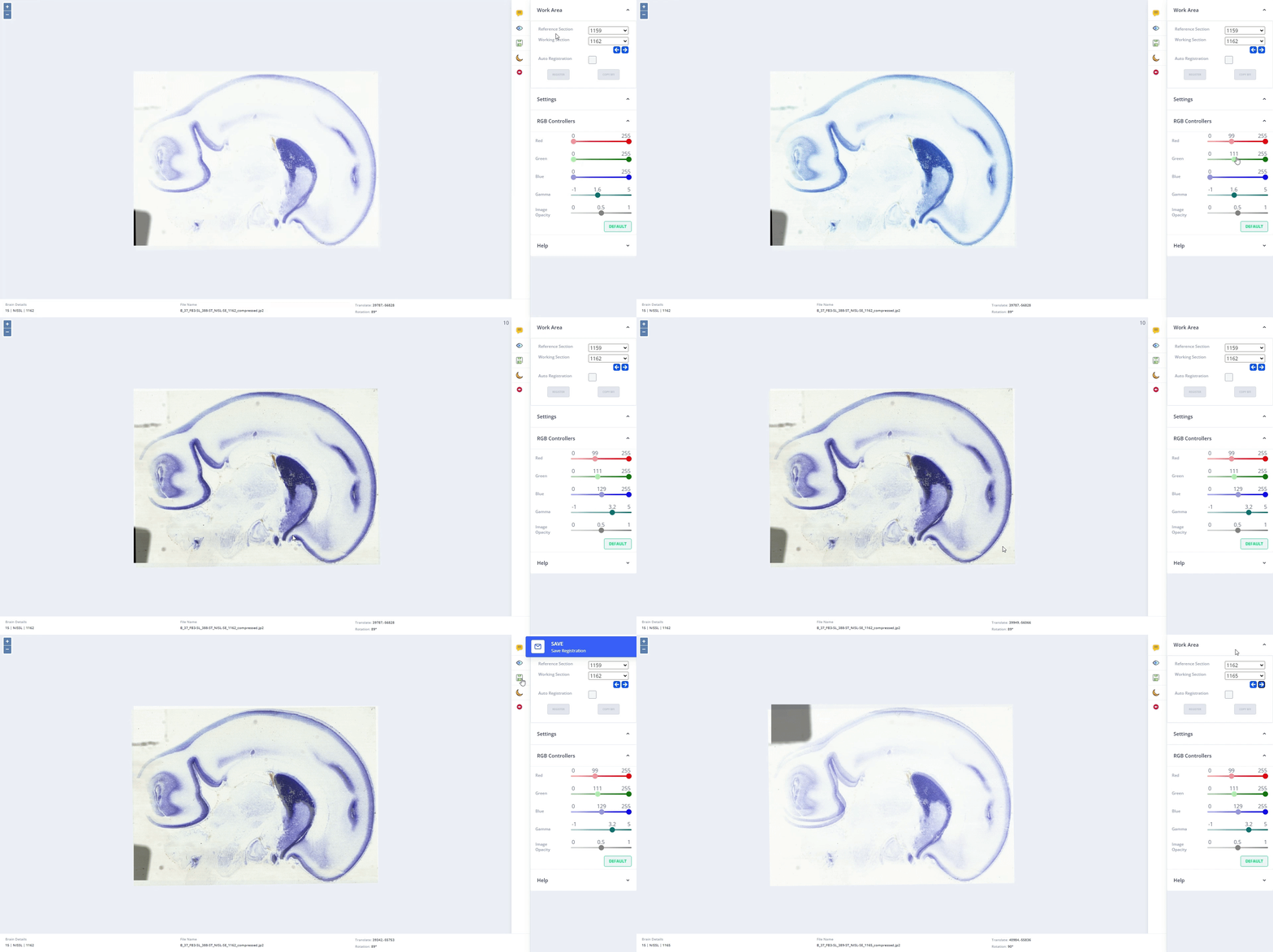
Registration (Manual & Automatic)
This tool aligns images from different modalities, time points, or subjects to a common coordinate system. It supports precise spatial correspondence for comparative analysis and integration.
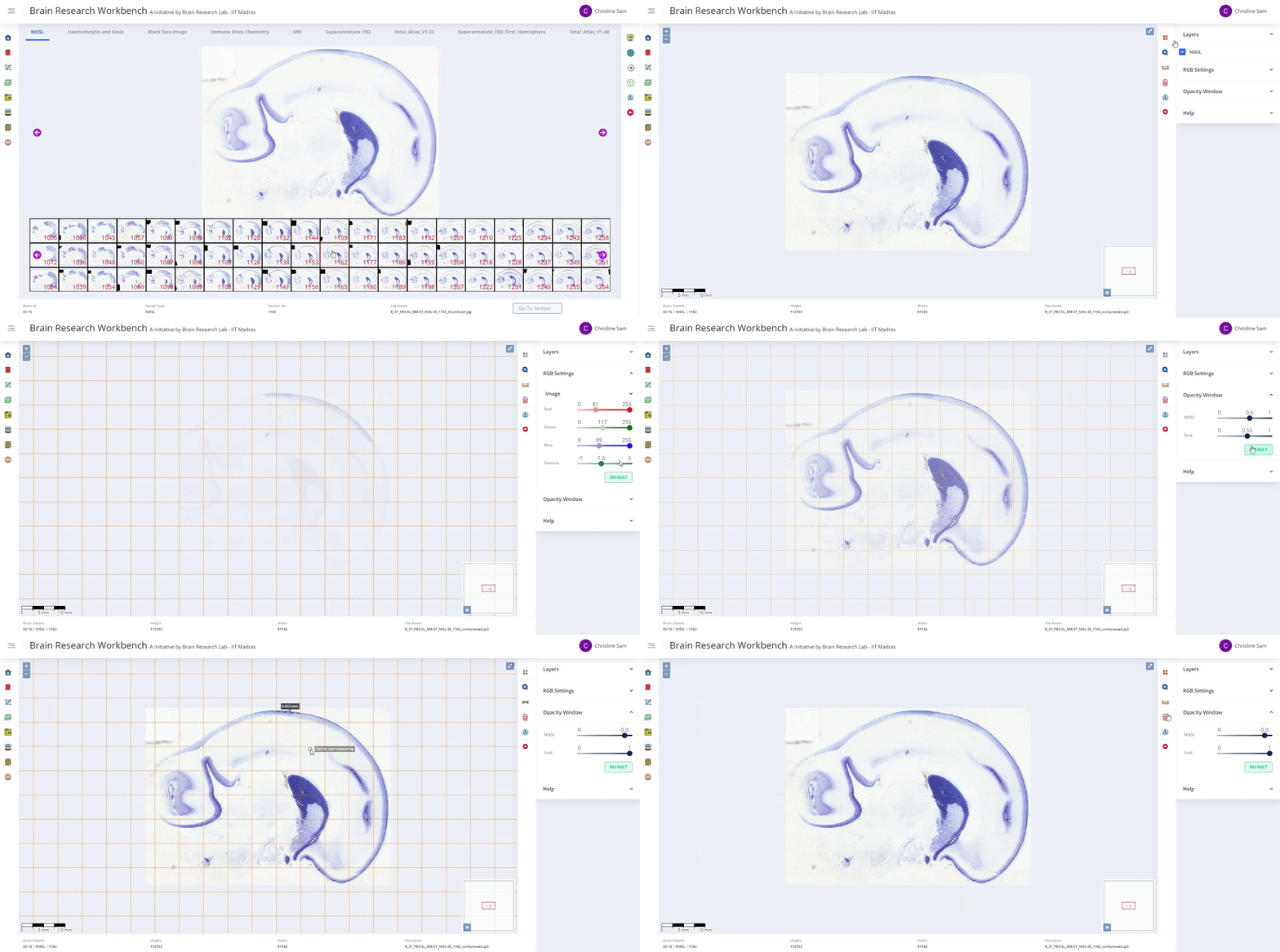
Series Viewer
This tool facilitates exploration and analysis of large image series. It enables interactive inspection for identifying patterns and trends across datasets.
Technology & Engineering
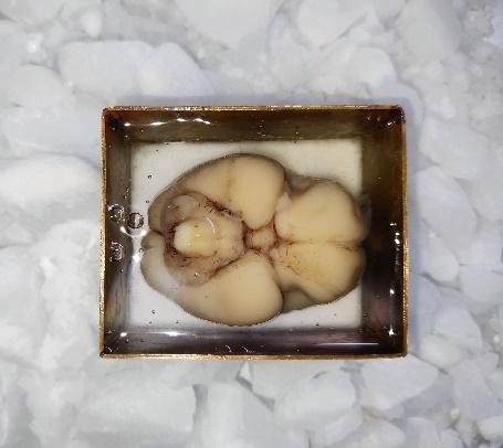
Brain Freezing
This technology platform enables freezing of large human brains in stereotaxic coordinates with minimal tissue damage and artifacts. It ensures preservation of anatomical integrity for downstream histological processing.
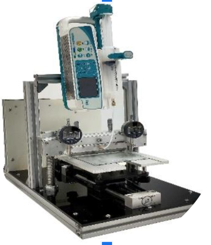
Slot Die Coater
This automated system applies a uniform 10–20 µm thin adhesive layer on glass slides. It preserves tissue integrity during tape-transfer cryo-sectioning.
Automated Tissue Transfer
This automated system performs cryo-sectioning and transfers tissue onto glass slides with minimal tissue damage. It ensures consistent and reliable section handling.
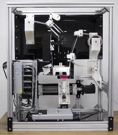
Block Face Imaging
This system serves as an intermediate step in the histopathology pipeline for imaging tissue blocks. It enables detection of structural and volumetric changes in the brain.
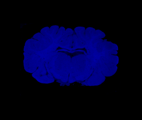
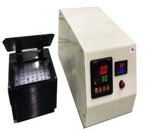
UV Curing Station
This system enables hands-free and tool-free transfer of 20 µm thick tissue sections onto glass slides. The transfer is achieved through controlled UV exposure.
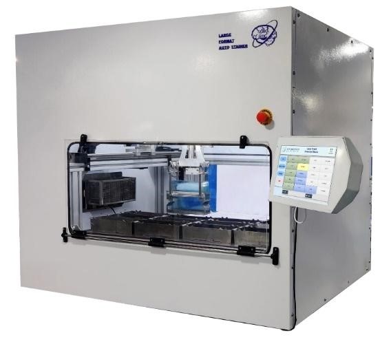
Large Format Autostainer
This custom-built automated staining system processes tissues mounted on multiformat slides (6×8, 5×7, 2×3, and 1×3). It ensures staining uniformity and consistency across formats.
Large Format Coverslipper (6×8)
This automated system performs uniform, bubble-free coverslipping of 6×8 slides. It supports a throughput of up to 30 slides per hour.
.jpg)
Automated Coverslipper (2×3)
This fully automated system performs coverslipping of 2×3 slides. It supports high-throughput operation of up to 50 slides per hour.
.jpg)
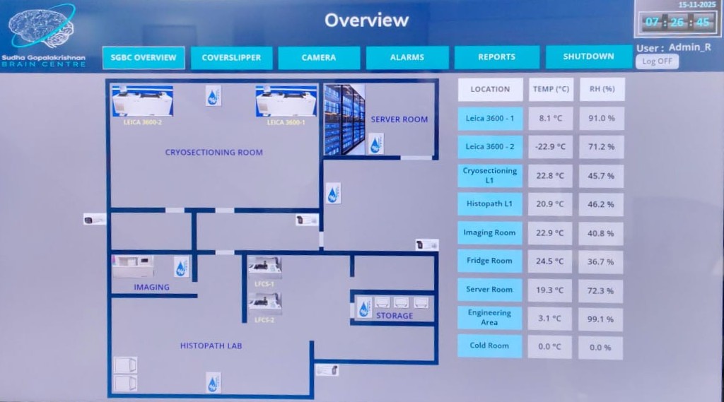
Monitoring Dashboard
This system monitors critical laboratory operations, including environmental parameters, coverslipping, and staining. It generates reports and alerts users during failures.
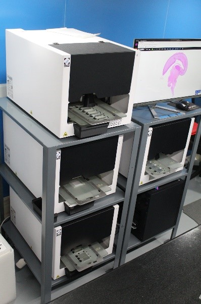
Scanning Station
These custom whole-slide scanners digitise multiformat slides at 0.5 µm per pixel resolution. They are developed in collaboration with an indigenous company.